 here's a lot of confusion out there about mRNA vaccines. Are they a form
of gene therapy? Is the mRNA vaccine dangerous? How does an mRNA molecule
induce the body to produce antibodies? Below are some answers.
Most people know that proteins can be toxic. Many viruses and bacteria produce toxic proteins. Diphtheria toxin, for example, is one of the most poisonous molecules in existence.
When a researcher wishes to do recombinant DNA research, they are required by their institutional biosafety committee to certify that they are not planning to produce or use any 'select agents'. This is a category reserved for the most dangerous substances on earth—think sarin, ricin, and the 1918 influenza virus—and putting them in an expression vector is widely agreed to be a Bad Idea.

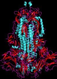
Spike protein in its normal 3D conformation. Structural biology rarely shows up in our movies or TV shows, but would make them much more interesting
Is Spike protein dangerous?
The Spike protein in SARS-CoV-2 isn't in that category, but it too is toxic. This should not be surprising: after all, proteins do the actual work of a cell, so they are responsible for much of its pathogenicity. However, it's not so easy to disentangle between direct toxic effects and indirect toxic effects caused by the immune system's reaction to it.
Both Pfizer/BioNTech and Moderna vaccines are, in effect, expression vectors for the full-length Spike protein, and so any concerns about Spike are also concerns about the mRNA vaccines. This does not necessarily mean the vaccines are toxic: toxicity depends on where the toxin is, how much of it is there, and how long it remains.
Nonetheless, the research literature is clear that Spike is indeed toxic. Robles et al.[1] found that Spike protein by itself, with no virus present, causes dysfunction of endothelial cells lining the blood vessels by causing a protein called NF-κB to translocate to the nucleus. NF-κB is a transcription factor that induces the expression of proinflammatory cytokines such as TNFα, interleukin-1β, and interleukin-6. Endothelial cell damage is also produced in comorbidities such as aging, obesity, and diabetes. The most harmful part of Spike seems to be the RGD tripeptide [2], which reproduces some of these features. RGD is not to be confused with RBD, the receptor binding domain.
Robles et al.[1] write:
Accumulating evidence has defined COVID-19 as a vascular disease. Blood vessel injury causes progressive lung damage and multi-organ failure in severe COVID-19, owing to edema, intravascular coagulation, vascular inflammation, and deregulated inflammatory cell infiltration.
Potential mechanisms of vascular dysfunction in COVID-19 include EC [endothelial cell] death in response to SARS-CoV-2 entry and replication, the binding of the spike protein of SARS-CoV-2 to the ACE2 receptor causing its downregulation and subsequent mitochondrial dysfunction, the activation of the proinflammatory kallikrein-bradykinin system, and the accumulation of proinflammatory and vasoconstrictor angiotensin II. In addition, the spike protein can trigger the expression of proinflammatory cytokines and chemokines, the production of toxic reactive oxygen species, and cell death.
Another paper [3] suggests that Spike may even be responsible for long-covid syndrome. The author suggests it could disrupt the blood-brain barrier, or BBB, by activating Toll-like receptors, which are pattern recognition receptors in the innate immune system.
Other reports [4,5] confirm that Spike protein crosses the BBB. The authors showed that intranasal infection induced ischemic-like reactivity in pericytes, which are cells that make up the BBB, and that the bare S (spike) protein, as well as intact viruses, readily crossed the BBB and entered the parenchymal brain space. It was even able to reach the hippocampus, which is essential for memory formation. S1, an activated fragment of S, crosses the BBB by a process called adsorptive transcytosis with the aid of ACE2, which is abundantly found on cells in the BBB.
The spike protein produces a variety of highly toxic effects on cultured pericytes, which are reproduced in live mice. Thus, even though the immune system, as always, gets its grubby little mitts on the matter, we have solid evidence that there is a direct toxic effect of the spike protein.
What's in those mRNA vaccines?
These mRNA vaccines aren't just made by covering the sequence for S with a tasty lipophilic coating and injecting it into people. The mRNA is given chemical and sequence modifications to make it survive longer and transcribe faster. Then it's slapped inside its tasty coating.
Xuhua Xia of the University of Ontario has a great article on this [6]. The vaccine, as I mentioned, is essentially an expression vector. Pfizer's BNT162b2 and Moderna's mRNA-1273 both contain a modified SARS-CoV-2 FLSP antigen, which is to say the full-length Spike protein.
Previous attempts to make vaccines against spike proteins in MERS-CoV, SARS-CoV, and similar coronaviruses resulted in serious side effects, including pulmonary eosinophilic infiltration (PEI) and antibody-dependent disease enhancement (ADDE), which occur when the antibody is taken up by the Fc receptor in macrophages [7] by phagocytosis.
The Fc, or ‘fragment crystallizable’ region, is the back end of an antibody opposite to the part that binds to the antibody's target. All antibodies have similar Fc regions. Macrophages and some other cells have receptors on their surface that bind to the Fc region of antibodies. When this happens, the antibody gets pulled into the cell by phagocytosis and the macrophage's protective mechanisms become activated. This is usually beneficial, but it can be harmful if the antibody is bad, as happens in autoimmune diseases.[8] If the antibody happens to be unable to neutralize the virus, this causes the receptor to act as a Trojan horse that can pull a live virus in with it, making a subsequent infection much worse. The classic case a few years ago was with Dengue virus antibodies, which turned ordinary cases of Dengue into Dengue hemorrhagic fever, a deadly disease found in patients who had been vaccinated or had a previous infection.
The other problem with mRNA is that it is rapidly destroyed by the cell, often only a few minutes after being produced. mRNA vaccines have to be carefully engineered to avoid these problems. Here's what they have done.
Two amino acids (KV or lysine-valine at 986 and 987) are replaced by PP (proline-proline) to stabilize Spike in its pre-fusion state, that is to say the 3D conformation of the Spike protein before it attaches to the cell. This mutated S protein is called S-2P.
In Pfizer's BNT162b2, all U (uridine) nucleotides are replaced by N1-methylpseudouridine (Ψ) to reduce degradation and reduce immune reaction to the RNA itself. This unnatural base ‘wobbles’, increasing the error rate. This can create problems: replacing a U with Ψ in a stop codon can cause the ribosome to read through the end of the mRNA, producing a longer protein with unknown function.[9] For this reason, the designers put two stop codons in BNT162b2 (UGA-UGA) and three in mRNA-1273 (UGA-UAA-UAG). However, since misreading can cause a frameshift, this strategy is not always 100% effective.
The vaccine is injected into muscle because the ZAP-mediated RNA degradation enzymes, which preferentially attack CpG motifs in RNA, are low in muscle, so muscle injection increases stability.
The vaccine's translation initiation signal is a modified Kozak sequence, which is designed to maximize expression. The 5′ and 3′ untranslated regions (UTRs) are based on human alpha-globin protein, which is highly expressed. This causes the cell to make more of it.
The codon usage is optimized for faster expression, and other features of mRNA such as its 5′ cap and poly(A) tail are modified to enhance its stability and expression efficiency in the cell. A mRNA molecule so modified can survive in a cell for as long as a week instead of just a few minutes.[10]* However, codon optimization can be detrimental, as slow translation (caused by rare codons) is necessary for some proteins to fold properly [11]. This isn't a big deal in the vaccine, as Spike is not intended to be a functional protein and it's going to be degraded anyway.
Is an mRNA vaccine gene therapy?
The mRNA vaccines are often said to be ‘generally safe’, but many people have become suspicious due to the overly rosy picture often painted in the media. A legitimate concern is the long history of side effects from vaccines, sometimes including paralysis, severe disease, and even death as alluded to above. These possibilities are downplayed in the scientific literature and ignored in the press because the authors wish to encourage people to get vaccinated. Skeptics see that they are being dishonestly manipulated and assume the worst.
However, the mRNA vaccines are most assuredly not gene therapy. Gene therapy is therapy that affects the genes, and it is by definition a permanent change. A gene is a DNA molecule. mRNA is not a gene and it does not go to the nucleus. mRNA has no effect on a cell's genes. Yes, the mRNA contains genetic information, but it is not incorporated into the genome, nor does the cell reverse-translate it into DNA. Calling it gene therapy is inaccurate.
How does a mRNA vaccine produce antibodies?
A vaccine expressing Spike produces large amounts of the protein inside the cell. But antibodies are in the bloodstream. How can the vaccine produce an antibody, which is an extracellular B-cell response, when the antigen is inside the cell? There are several possible ways:

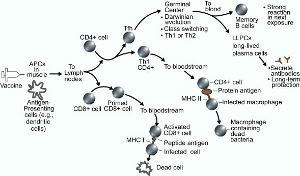
Simplified diagram of adaptive immune system after vaccination
The Spike protein created in the cell is gradually broken down into peptides. These peptides are taken to the surface and presented to the outside by MHC class I proteins (in humans these are also known as HLAs or human leukocyte antigens) on the surface. This is the cell's way of telling the immune system what's going on inside the cell. If the immune system finds an alien protein, cytotoxic CD8+ T cells attack and kill the cell. When the cell lyses, some of those peptides are released to the extracellular space.
More common is when the mRNA enters immune cells such as dendritic cells, which are immune cells scattered throughout the body. In this case, Toll-like receptor TLR3 (which recognizes double-stranded RNA), and TLR7 and TLR8 (which recognize single-stranded RNA) induce the secretion of interferon, some of which makes its way into ordinary non-immune cells, and through a very complicated pathway activates the cell's own interferon response, which inhibits viral mRNA translation.
Inflammation increases the rate of entry of blood plasma into inflamed tissue and helps the body carry antigens back to the lymphoid tissues where antibodies are made.
When dendritic cells containing the antigen travel to the lymph nodes, they can prime CD4 and CD8 T lymphocytes. The CD8 cells are cytotoxic, which means their function is to kill infected cells. The CD4 cells, once primed by the dendritic cells, change into Th1 cells or Tfh (T follicular helper) cells. The Tfh cells initiate a germinal center (GC) reaction, which results in the maturation of B cells in the lymph nodes. These are called affinity-matured memory B cells and, along with long-lived plasma cells, they manufacture and secrete antibodies.[12] Antibody maturation sounds complicated, and it is, but it is also a fascinating process of rapid cell-based directed Darwinian evolution which is briefly described here.
If the vaccine's coding sequence contained a secretion signal (which as far as I know none of them do), the expressed Spike protein would pass through the membrane into the extracellular space. If the mRNA contained a routing sequence, the protein would also be routed to MHC class II loading compartments in immune cells, thereby inducing a more potent and sustainable response. This would be a good way of enhancing the B-cell response, but it would also expose the patient to more of the toxic spike protein.
Covid-19 is now recognized as a vascular disease.[1] If some of the vaccine reaches the bloodstream, it can induce toxic effects on endothelial cells and thereby activate innate immune responses throughout the cardiovascular system.
Much of the confusion comes from a sort of nannyism on the part of the science media and scientific authorities. These days, people readily notice when overly-optimistic information is being spoon-fed to them. I've seen it myself in countless scientific articles, news stories, and blog posts. There is no better way to generate skepticism and cynical over-reaction. No human activity is without risk. Pretending something is perfectly safe when it is clearly not only convinces people you're hiding something. If you want to know why there is vaccine misinformation, there's the reason.
* Update (Feb 26 2022): A recent paper [13] found that mRNA and spike antigen from vax remain in dendritic cells and B cells in germinal centers of lymphatic tissue not just for a week, but for up to 60 days after the second dose of vaccination.
1. Robles JP, Zamora M, Adan-Castro E, Siqueiros-Marquez L, Martinez de la Escalera G, Clapp C. The spike protein of SARS-CoV-2 induces endothelial inflammation through integrin a5β1 and NF-κB signaling. J Biol Chem. 2022 Feb 7:101695. doi: 10.1016/j.jbc.2022.101695. PMID: 35143839; PMCID: PMC8820157.
2. Beaudoin CA, Hamaia SW, Huang CL, Blundell TL, Jackson AP. Can the SARS-CoV-2 Spike Protein Bind Integrins Independent of the RGD Sequence? Front Cell Infect Microbiol. 2021 Nov 18;11:765300. doi: 10.3389/fcimb.2021.765300. PMID: 34869067; PMCID: PMC8637727.
3. Theoharides TC. Could SARS-CoV-2 Spike Protein Be Responsible for Long-COVID Syndrome? Mol Neurobiol. 2022 Jan 13:1–12. doi: 10.1007/s12035-021-02696-0. PMID: 35028901; PMCID: PMC8757925.
4. Rhea EM, Logsdon AF, Hansen KM, Williams LM, Reed MJ, Baumann KK, Holden SJ, Raber J, Banks WA, Erickson MA. The S1 protein of SARS-CoV-2 crosses the blood-brain barrier in mice. Nat Neurosci. 2021 Mar;24(3):368–378. doi: 10.1038/s41593-020-00771-8. PMID: 33328624; PMCID: PMC8793077.
5. Khaddaj-Mallat R, Aldib N, Bernard M, Paquette AS, Ferreira A, Lecordier S, Saghatelyan A, Flamand L, ElAli A. SARS-CoV-2 deregulates the vascular and immune functions of brain pericytes via Spike protein. Neurobiol Dis. 2021 Dec;161:105561. doi: 10.1016/j.nbd.2021.105561. PMID: 34780863; PMCID: PMC8590447.
6. Xia X. Detailed Dissection and Critical Evaluation of the Pfizer/BioNTech and Moderna mRNA Vaccines. Vaccines (Basel). 2021 Jul 3;9(7):734. doi: 10.3390/vaccines9070734. PMID: 34358150; PMCID: PMC8310186.
7. Malik JA, Mulla AH, Farooqi T, Pottoo FH, Anwar S, Rengasamy KRR. Targets and strategies for vaccine development against SARS-CoV-2. Biomed Pharmacother. 2021 May;137:111254. doi: 10.1016/j.biopha.2021.111254. PMID: 33550049; PMCID: PMC7843096.
8. Ben Mkaddem S, Benhamou M, Monteiro RC. Understanding Fc Receptor Involvement in Inflammatory Diseases: From Mechanisms to New Therapeutic Tools. Front Immunol. 2019 Apr 12;10:811. doi: 10.3389/fimmu.2019.00811. PMID: 31057544; PMCID: PMC6481281. Link
9. Adachi H, De Zoysa MD, Yu YT. Post-transcriptional pseudouridylation in mRNA as well as in some major types ofnoncoding RNAs. Biochim. Biophys. Acta Gene Regul Mech. 2019,1862, 230–239.
10. Sahin U, Karikó K, Türeci Ö. mRNA-based therapeutics: developing a new class of drugs. Nat. Rev. Drug Discov. 2014;13(10):759–780.
11. Kimchi-Sarfaty C, Oh JM, Kim IW, Sauna ZE, Calcagno AM, Ambudkar SV,
Gottesman MM. A "silent" polymorphism in the MDR1 gene changes substrate
specificity. Science. 2007 Jan 26;315(5811):525–528.
doi: 10.1126/science.1135308.
Erratum in: Science. 2007 Nov 30;318(5855):1382-3.
Erratum in: Science. 2011 Oct 7;334(6052):39. PMID: 17185560.
12. Bettini E, Locci M. SARS-CoV-2 mRNA Vaccines: Immunological Mechanism and Beyond. Vaccines (Basel). 2021 Feb 12;9(2):147. doi: 10.3390/vaccines9020147. PMID: 33673048; PMCID: PMC7918810.
13. Röltgen K, Nielsen SCA, Silva O, Younes SF, Zaslavsky M, Costales C, Yang F, Wirz OF, Solis D, Hoh RA, Wang A, Arunachalam PS, Colburg D, Zhao S, Haraguchi E, Lee AS, Shah MM, Manohar M, Chang I, Gao F, Mallajosyula V, Li C, Liu J, Shoura MJ, Sindher SB, Parsons E, Dashdorj NJ, Dashdorj ND, Monroe R, Serrano GE, Beach TG, Chinthrajah RS, Charville GW, Wilbur JL, Wohlstadter JN, Davis MM, Pulendran B, Troxell ML, Sigal GB, Natkunam Y, Pinsky BA, Nadeau KC, Boyd SD. Immune imprinting, breadth of variant recognition, and germinal center response in human SARS-CoV-2 infection and vaccination. Cell. 2022 Jan 25:S0092-8674(22)00076-9. doi: 10.1016/j.cell.2022.01.018. PMID: 35148837; PMCID: PMC8786601. Link
feb 18 2022, 6:25 am. updated feb 19 2022. diagram added feb 19, 6:06 am
